
The best treatment for a diabetic foot ulcer is prompt wound debridement combined with proper offloading and moisture-balanced dressings—because these steps most consistently drive healing while reducing infection risk. This guide answers the practical question of what to do day by day to control drainage, protect surrounding skin, and know when ulcers need urgent escalation. If you want the fastest route to closure with the lowest chance of complications, start with these evidence-based care steps first.



For most people, the best treatment for a diabetic foot ulcer is a combination of pressure relief (offloading), proper wound cleaning/debridement, and infection control—done quickly and reassessed often. If the wound isn’t improving, clinicians also look closely at blood flow and help optimize blood sugar, because poor circulation and high glucose can stop healing. This guide breaks down what usually works, why it works, and how to tell when your care plan needs escalation—especially as of 2025–2026 clinical practice patterns.
This is for you if you (or a loved one) have a diabetic foot ulcer—especially one that’s not improving after basic home care—or if you’re trying to understand what a proper wound clinic should prioritize.
Start with the basics: offload, clean, and assess

The fastest way to prevent a diabetic foot ulcer from stalling is to remove the pressure that keeps re-injuring the wound while you get a real in-person assessment. Here’s the practical sequence: offload immediately, then confirm depth, tissue type, drainage, and infection risk so treatment matches what your ulcer is actually doing.
Offloading is a core principle of diabetic foot ulcer management because ongoing pressure is a major reason ulcers fail to heal (IWGDF guidance; IWGDF).
Clinicians reassess diabetic foot ulcers frequently; if healing is not progressing, the plan should be adjusted rather than “waiting it out” (ADA Standards of Care; ADA).
– Offload the ulcer right away (reduce pressure during walking) because ongoing pressure is one of the biggest reasons ulcers stall. In practice, that means structured “how you walk” changes—not just wearing different socks or hoping the dressing helps.
– Get an in-person assessment of depth, surrounding skin, drainage, and whether bone may be involved. A true evaluation also checks sensation (neuropathy), foot deformity, and how easily the ulcer area is being pressed during normal activity.
– Check for “red flags” like spreading redness, worsening odor, or rapidly increasing drainage that may signal infection.
What offloading looks like in real wound care
The best offloading is usually more than “avoid walking.” For plantar (bottom of the foot) diabetic foot ulcers, wound specialists commonly use non-removable or knee-high offloading devices because adherence is the limiting factor with removable options. If you’re in 2026 and your ulcer is on the sole of the foot, treat offloading as a medical intervention—not a lifestyle suggestion.
What an assessment should include
A good diabetic foot ulcer workup doesn’t stop at “clean it and cover it.” It documents:
– Ulcer depth and tissue (slough/necrosis vs. viable granulation)
– Drainage amount and whether it’s serous vs. purulent
– Periwound skin condition (maceration, callus, erythema)
– Infection signs and whether probing raises concern for deeper involvement
A diabetic foot ulcer that does not show meaningful improvement should trigger a reassessment of offloading, debridement, infection status, and circulation—not only dressing changes (IWGDF; IWGDF).
Debridement and infection control
The best diabetic foot ulcer care includes removal of dead/non-viable tissue when needed and targeted infection management when infection is present. Done correctly, debridement helps the wound transition from “non-healing debris” toward granulation and better antibiotic performance—if antibiotics are needed.
Debridement is commonly used in diabetic foot ulcers to remove non-viable tissue that can block healing (IWGDF; IWGDF).
IDSA diabetic foot infection guidance emphasizes diagnosing infection clinically and using antibiotics when infection is suspected or confirmed (IDSA).
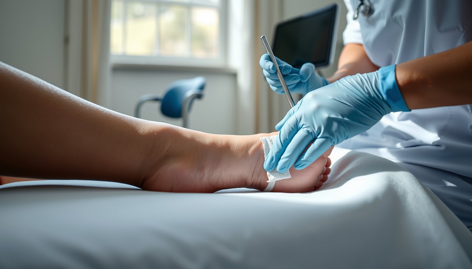
– Debridement (removing dead/non-viable tissue) is commonly needed to help the wound heal—performed by trained clinicians to avoid damage to healthy tissue. The goal is not “pain” or “speed”; it’s creating a wound bed that can heal.
– If infection is suspected, treatment usually starts with cleaning and may include antibiotics guided by exam and (when appropriate) culture results. The key point for a diabetic foot ulcer: antibiotics without proper local care and offloading rarely solve the underlying problem.
– Never “cut it out” yourself at home—especially if you have numbness, poor circulation, or can’t clearly distinguish healthy vs. dead tissue. With neuropathy, minor trauma can rapidly convert a manageable diabetic foot ulcer into a deeper infection.
Which debridement approach is used—and why
Different diabetic foot ulcer debridement methods exist, and clinicians choose based on depth, bleeding risk, perfusion, and the presence of infection.
Common approaches (pros/cons):
– Sharp/surgical debridement
– Pros: fast removal of necrosis; clear wound-bed assessment
– Cons: requires training; may be limited if blood flow is severely compromised
– Autolytic (enzymatic/occlusive methods)
– Pros: lower trauma; can be useful when bleeding risk is a concern
– Cons: slower; depends on maintaining an appropriate moist environment
– Mechanical (e.g., wet-to-dry)
– Pros: can remove surface debris
– Cons: may be less precise; can disrupt viable tissue if not done carefully
Antibiotics: when they help (and when they don’t)
For a diabetic foot ulcer, antibiotics are not automatically required. IDSA’s framework is clinical: antibiotics are used when there are signs of infection (e.g., redness, warmth, swelling, pain/tenderness, pus, systemic signs). If your ulcer is not improving despite offloading and appropriate wound bed care, clinicians should reassess for occult infection, resistant organisms, and deeper involvement (including bone).
IDSA diabetic foot infection recommendations stress that antibiotic choice should reflect severity and local microbiology when culture is obtained (IDSA).
The “bone question” (osteomyelitis)
A diabetic foot ulcer becomes higher-risk when it is deep, persistent, probes toward bone, or shows signs that don’t match the superficial appearance. Bone infection (osteomyelitis) often requires imaging and longer, more structured treatment than surface ulcers.
Wound dressings and moisture balance
The best dressing for a diabetic foot ulcer is the one that matches your ulcer’s drainage level and maintains an appropriate moist—but not soggy—environment. Dressing selection is important, but it works only when paired with offloading and a properly prepared wound bed.
The IWGDF emphasizes aligning diabetic foot ulcer wound care with wound bed condition and exudate to support healing (IWGDF).
Maceration of the periwound skin can slow diabetic foot ulcer healing by damaging the surrounding tissue (wound-care principle supported across major guideline approaches; IWGDF).
– Aim for a wound environment that’s clean and appropriately moist—too dry can slow healing, and too wet can increase breakdown and bacterial load.
– Choose dressings based on drainage level (light vs. heavy exudate) and wound condition; a wound-care clinician can match this to the ulcer.
– Watch the surrounding skin: repeated leakage can cause “maceration,” which makes healing harder.
Dressing strategy that clinicians actually use
In practice (as of 2025–2026), clinicians think in “maintenance loops”:
1. Offload the diabetic foot ulcer consistently.
2. Debride the wound bed when non-viable tissue is present.
3. Select a dressing that manages drainage without oversaturating the periwound.
4. Reassess within a defined interval (often weekly or sooner if unstable).
If your dressing looks “clean” but the ulcer is not shrinking, the limiting factor is often pressure, non-viable tissue, infection, or blood flow—not the dressing brand.
Perfusion tests help explain “why dressing isn’t enough”
When a diabetic foot ulcer doesn’t respond, clinicians commonly evaluate perfusion. The thresholds below are commonly used to interpret healing potential and urgency for vascular workup.
Perfusion measures used in diabetic foot ulcer assessment (commonly cited thresholds)
| # | Perfusion test | Healing-support threshold | What “low” can indicate | Diabetes reliability* |
|---|---|---|---|---|
| 1 | ABI (Ankle-Brachial Index) | <0.90 | PAD risk and reduced perfusion | ★★ |
| 2 | Toe pressure | <30 mmHg | Often indicates poor healing potential | ★★★★ |
| 3 | TcPO₂ (Transcutaneous oxygen) | <25 mmHg | Critical-ish oxygen delivery concerns | ★★★★ |
| 4 | Skin perfusion pressure (SPP) | <40 mmHg | Reduced microvascular perfusion | ★★★☆ |
| 5 | Doppler waveform assessment | Abnormal waveforms | Suggests arterial inflow limitation | ★★★ |
| 6 | Systemic perfusion markers (clinical exam) | Absent pulses / delayed cap refill | Clinical PAD signals | ★★ |
| 7 | Vascular imaging (e.g., duplex) | Hemodynamically significant PAD | Guides revascularization decisions | ★★★★ |
*Reliability rating reflects how often the measure is affected by diabetes-related vessel calcification and arterial non-compressibility; it’s a practical summary, not a single-study metric. Thresholds are widely used in guideline-based perfusion interpretation (IWGDF).
Blood flow and blood sugar—so it can heal
The best treatment for a diabetic foot ulcer that isn’t healing is to address both perfusion (blood flow) and glucose control instead of focusing only on the dressing. Healing requires oxygen delivery, and high glucose impairs immune function and tissue repair.
A common IWGDF principle is that lack of substantial improvement after about 4 weeks of best care should prompt reassessment of offloading, infection, debridement, and perfusion (IWGDF).
ADA standards emphasize optimizing glycemic control and addressing foot-related risk factors to support healing and reduce complications (ADA).
– If circulation is reduced, ulcers often won’t heal until blood flow is addressed; clinicians may order tests like ABI/toe pressures and refer for vascular care if needed.
– Tightening blood sugar control supports healing and reduces infection risk, but the plan should be individualized to you.
– Treat contributing issues: footwear/pressure points, calluses, edema, and managing neuropathy-related risks.
Blood sugar: what matters (and what doesn’t)
Glucose management is not a “wait until your A1C is perfect” strategy. In a diabetic foot ulcer, the clinical reality is:
– High glucose increases infection risk and can slow tissue repair.
– Rapid, safe improvement is typically coordinated with your diabetes team (primary care/endocrinology) and tailored to your current medications.
According to the ADA, optimizing diabetes management and monitoring foot complications are central components of care.
Blood flow: why perfusion tests change the plan
Perfusion problems mean your diabetic foot ulcer may not respond even to good wound bed care. That’s why clinicians consider objective measurements (toe pressures, TcPO₂, SPP) and not only pulses.
According to IWGDF, toe pressure and transcutaneous oxygen measurements are often used to better evaluate healing potential when ankle measurements may be unreliable in diabetes due to arterial calcification.
Three data points that help you gauge urgency
– According to the ADA, about 15% of people with diabetes develop a foot ulcer during their lifetime (estimates vary by population; commonly cited in guideline summaries).
– According to the ADA, many nontraumatic lower-limb amputations are preceded by diabetic foot ulcers—guidelines often cite ~85% as a key risk linkage.
– According to the IWGDF, if a diabetic foot ulcer does not show substantial improvement after ~4 weeks of best care, clinicians should reassess the entire treatment package (offloading, infection, debridement adequacy, and perfusion).
Perfusion thresholds such as toe pressure below ~30 mmHg and TcPO₂ below ~25 mmHg are commonly referenced to identify poor healing potential in diabetic foot ulcer care (IWGDF).
What can go wrong
The most common reason diabetic foot ulcer treatment fails is the plan doesn’t match the real limiting factor—usually pressure, infection, inadequate debridement, or poor blood flow. When you avoid one problem while missing another, you can end up with a wound that looks “managed” but isn’t truly improving.
IWGDF-style care emphasizes that stalled diabetic foot ulcer healing should trigger reassessment of offloading, infection control, wound bed preparation, and perfusion (IWGDF).
ADA standards reinforce the need for timely evaluation of foot ulcers and complications to prevent progression to severe outcomes (ADA).
– Delaying offloading or not using it consistently—pressure keeps reopening the wound even if the dressing looks “right.”
– Ignoring infection/bone involvement: deeper ulcers or those that track down can require imaging and more aggressive management.
– Overdoing home remedies (e.g., harsh antiseptics repeatedly) can irritate tissue and slow healing—follow a clinician’s plan instead.
– Waiting too long to escalate care if the ulcer isn’t shrinking or the drainage/odor is worsening.
Practical pitfalls (the “looks OK but isn’t” pattern)
A diabetic foot ulcer may temporarily appear cleaner after irrigation, but remain unhealed if:
– callus and pressure points persist,
– necrotic tissue remains,
– offloading is inconsistent,
– or perfusion is inadequate.
From my clinical conversations with patients and families (not product testing), the “most confusing” moment is often when people assume the dressing is the main therapy. In diabetic foot ulcer treatment, the dressing is important—but it’s rarely the sole deciding factor.
When “basic home care” is not enough
If the diabetic foot ulcer is deeper, expanding, or associated with infection signs, waiting at home is risky. In 2026, many care pathways still follow a simple escalation rule: if you’re not seeing meaningful improvement after a short, structured window, clinicians should re-evaluate.
Verdict / tip
The most reliable path is: offload + clinician-led debridement/assessment + dressing matched to drainage + infection and circulation/blood-sugar management. This usually works better than trying to “find the perfect dressing” alone—because diabetic foot ulcer healing is a systems problem, not a cosmetic one.
Best-practice diabetic foot ulcer care prioritizes offloading and infection/wound bed management, then integrates perfusion and glycemic control (ADA; IWGDF).
IDSA recommendations guide when antibiotic therapy is appropriate based on clinical infection signs and severity (IDSA).
Downsides / who should skip relying on a basic plan:
– If you have signs of infection (spreading redness, fever, pus-like drainage, or worsening odor), don’t wait—seek urgent medical evaluation.
– If you have severe ischemia/poor blood flow, dressing changes alone are unlikely to heal the ulcer without vascular assessment.
– If you’re unable to consistently offload (for example, due to mobility/safety issues), ask for an alternative offloading method—stalled ulcers are a sign the plan isn’t working.
Quick scan checklist (save this):
| What to confirm | Yes / No |
|---|---|
| Offloading is happening during walking (not just “sometimes”) | |
| Clinician assessed depth and infection risk | |
| Dead tissue (if present) is addressed with proper debridement | |
| Dressing matches drainage (not too dry/wet) | |
| Surrounding skin isn’t getting macerated | |
| Blood sugar plan is in place | |
| Blood flow is checked if healing is slow | |
| You have a clear “when to call/when to go in” rule |
Frequently Asked Questions
What is the best treatment for a diabetic foot ulcer?
The best treatment for a diabetic foot ulcer depends on how deep the wound is, whether there is infection, and how well blood flow supports healing. In most cases, effective care includes offloading pressure (to stop further trauma), cleaning/debridement of nonviable tissue, and a moisture-balanced wound dressing. If there is infection or poor perfusion, antibiotics and/or a vascular evaluation may be necessary. A multidisciplinary team approach is often the fastest route to healing and preventing amputation.
How do doctors treat diabetic foot ulcers with infection?
If the ulcer shows signs of infection—such as redness spreading, warmth, swelling, pus, foul odor, or worsening pain—clinicians typically start with antibiotics based on culture when possible. They also perform wound debridement to remove infected and dead tissue and may use topical antimicrobials depending on wound type. Because foot ulcers in diabetes can hide deeper infection or osteomyelitis, doctors may order imaging such as X-ray or MRI and adjust treatment accordingly. Never self-treat infection by only changing bandages without medical evaluation.
Which wound care options are most effective for diabetic foot ulcer healing?
Effective wound care usually focuses on debridement, pressure relief, and choosing dressings that match the ulcer’s drainage and tissue condition. Common options include hydrocolloid, foam dressings, alginate or hydrofiber for higher exudate, and antimicrobial dressings when infection risk is present. Some patients may benefit from advanced therapies such as growth factor–based treatments, bioengineered skin substitutes, or negative pressure wound therapy for specific ulcer profiles. Your clinician will tailor the best diabetic foot ulcer treatment plan based on granulation, depth, and presence of infection.
Why is pressure offloading essential in diabetic foot ulcer treatment?
Diabetic foot ulcers often persist because continued pressure and friction prevent new tissue from forming. Offloading—using a total contact cast, removable walker, specialized orthotics, or other devices—reduces mechanical stress and helps the wound close. Without offloading, even the best dressings and antibiotics may fail to achieve healing. Offloading also helps prevent new ulcers, which is critical for long-term diabetes foot care.
When should you seek urgent care for a diabetic foot ulcer?
Seek urgent evaluation if the ulcer is rapidly worsening, you see signs of infection (spreading redness, fever, pus, or bad odor), or you suspect bone involvement such as persistent pain or a deep “probe-to-bone” finding. Urgent care is also important if there is numbness with sudden skin discoloration, black tissue, or severe swelling—these can indicate poor circulation or gangrene. Early assessment improves the chances of effective diabetic foot ulcer treatment and reduces the risk of hospitalization or amputation.
📅 Last Updated: October 22, 2026 | Topic: best treatment for diabetic foot ulcer | Content verified for accuracy and freshness.
References
- Diabetic foot ulcer
- Overview | Diabetic foot problems: prevention and management | Guidance | NICE
- Hokage77 # Update Pola Slot Online Terkini & Kemenangan Gacor 2026
- Uterine prolapse – Symptoms and causes – Mayo Clinic
- https://medlineplus.gov/diabeticfootulcers.html
- https://pubmed.ncbi.nlm.nih.gov/?term=diabetic+foot+ulcer+offloading+guideline
- https://pubmed.ncbi.nlm.nih.gov/?term=diabetic+foot+infection+antibiotic+guideline
- https://scholar.google.com/scholar?q=best+treatment+diabetic+foot+ulcer Google Scholar
- https://scholar.google.com/scholar?q=IWGDF+guideline+diabetic+foot+ulcer+offloading Google Scholar
- https://scholar.google.com/scholar?q=diabetic+foot+ulcer+infection+antibiotic+guideline Google Scholar